Journal of JCIC

Online edition: ISSN 2432–2342
JCIC学会事務局 JCIC学会事務局
〒162-0801東京都新宿区山吹町358-5アカデミーセンター Academy Center, 358-5 Yamabuki-cho, Shinju-ku, Tokyo 162-0801, Japan
Journal of JPIC 2(1): 14-20 (2017)
doi:10.20599/jjpic.2.14

原著Original Article

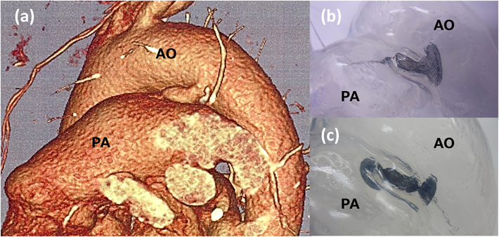
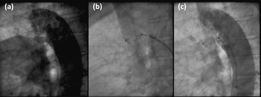
患者データから自施設で作製した中空立体模型を用いた,成人動脈管開存の経皮的閉鎖術シミュレーションSimulation using patient-specific hollow three-dimensional models for percutaneous closure of patent ductus arteriosus in adult

1自治医科大学とちぎ子ども医療センター小児手術・集中治療部Pediatric Operating Suite and Intensive Care Unit, Jichi Children’s Medical Center Tochigi ◇ Tochigi, Japan

2自治医科大学小児科Department of Pediatrics, Jichi Medical University ◇ Tochigi, Japan

3自治医科大学成人先天性心疾患センターAdult Congenital Heart Disease Center, Jichi Medical University ◇ Tochigi, Japan

4自治医科大学とちぎ子ども医療センター小児・先天性心臓血管外科Pediatric and Congenital Cardiovascular Surgery, Jichi Children’s Medical Center Tochigi ◇ Tochigi, Japan

受付日:2017年6月12日Received: June 12, 2017
受理日:2017年7月6日Accepted: July 6, 2017
発行日:2017年8月31日Published: August 31, 2017
HTMLPDFEPUB3

背景と目的】成人動脈管開存(PDA)の経皮的閉鎖術において,留置時の閉鎖栓の形状を正確に予測することは困難である.患者データから作製した中空立体模型を用いた,成人PDAの経皮的閉鎖術シミュレーションの有用性を検討する.

対象と方法】対象は成人PDA4症例.年齢は54~75歳,Krichenko分類はE 3症例,D1症例であった.CT画像データから3Dプリンターで造形した実体模型を元に透明シリコーン製中空模型を作製し,AMPLATZER™ Duct OccluderおよびAMPLATZER™ Vascular Plug IIの留置形状をシミュレーションして閉鎖術に臨んだ.

結果】中空模型の作製時間は3日,作製費用は約1万円であった.留置した閉鎖栓は良好に透見でき,手にとって様々な角度から観察することで,最適な閉鎖栓を選択しえた.

考察と結論】透明シリコーン製中空立体模型はPDAの経皮的閉鎖術シミュレーターとして優れ,特に成人症例で有用性が高い.

Background and Objective: It is challenging to accurately predict the shape and stability of occlusion devices during percutaneous closure of patent ductus arteriosus (PDA) in adults. In this study, we evaluate the utility of percutaneous closure simulation of PDA in adults using patient-specific hollow three-dimensional (3D) models created from computed tomography data.

Methods: The study was conducted on four adult patients with PDA, whose age ranged from 54 to 75 years. While three were type E as per the Krichenko classification, one was type D. We created a transparent silicone hollow model using a solid model, which was printed by a personal 3D printer from contrast-enhanced multidetector computed tomography data, as a mold. All processes were carried out at our institution. Before actually performing percutaneous closure of PDA, we simulated procedures using these models and investigated the shape and stability of AMPLATZER™ Duct Occluder/AMPLATZER™ Vascular Plug deployed in them.

Results: Briefly, the time and cost required to create a hollow 3D model were about 3 days and 10,000 Japanese Yen, respectively. It was very easy to recognize the shape and stability of deployed devices in these models because the devices were clearly visible from outside. In fact, these models could be picked up by hands and observed from various angles, which was helpful in selecting optimum devices for closure.

Conclusions: The transparent silicone hollow models are an excellent simulator of percutaneous closure of PDA, especially in adults.

Key words: simulation; three-dimensional printing; patent ductus arteriosus; percutaneous closure; adult

はじめに

近年,3Dプリント技術の発達は目覚ましく,医療分野にも応用されつつある.本邦においても脳神経外科,整形外科領域では「画像等手技支援加算」が保険収載されており,既に実用化されている.先天性心疾患診療分野でも,3Dプリントされた立体模型を用いた治療シミュレーションの報告がみられるようになってきた1–8).しかし,個別の患者データを基にした立体模型の作製は,主として専門業者に依頼し高額である3)ことなどから,いまだ日常診療への普及は進んでいない9)

成人における動脈管開存(PDA)では,動脈管は小児に比べ太く長く,血管壁の石灰化10, 11)や瘤形成を伴うことが多い12).経皮的閉鎖術に際しては,血管造影やCT画像などを基に閉鎖栓を選択する12, 13)が,本邦では使用可能なデバイスが限られるうえ,留置時の閉鎖栓形状を正確に予測することは困難で,経験に頼らざるをえない.

我々は,市販のパーソナル3Dプリンターを用いて,患者データからシリコーン製中空立体模型を自施設で作製する簡易な手法を確立した.本研究の目的は,この手法で作製した模型を用いた,成人PDAの経皮的閉鎖術シミュレーションの有用性を検討することである.

対象と方法

1. 対象とシミュレーションの方法

対象は成人PDA 4症例で,年齢は54~75歳,全て女性であった(Table 1).当施設では成人PDA症例に対し,血管形態把握の目的で原則的全例に術前の造影CT検査を施行している.他施設でCT検査を施行された1症例を除く3症例で,十分な説明の後,文書によるCT検査の同意を得た.対象の動脈管形態はKrichenko分類14)のEないしDで,CT画像で計測した動脈管の最小径は4.1~5.2 mm,長さは18.1~26.0 mmで,2症例が石灰化を伴っていた.肺/体血流比は1.4~2.4で,肺高血圧合併症例はなかった.全例NYHA分類のII度であった.同意を得たうえで患者のCT画像データを用いて,後述の方法で透明シリコーン製中空模型を作製し,AMPLATZER™ Duct Occluder(ADO: St. Jude Medical, St. Paul, MN, USA)およびAMPLATZER™ Vascular Plug II(AVP: St. Jude Medical, St. Paul, MN, USA)の留置形状をシミュレーションした後,経皮的閉鎖術に臨んだ.ADOは肺動脈側から,AVPは大動脈側からアプローチして留置した.PDA閉鎖にAVPを使用するにあたっては,院内倫理委員会の承認を得た.経皮的閉鎖術の際には模型をカテーテル検査室に持ち込んで,シミュレーションでの閉鎖栓の留置形状を参照した.疾患と治療の理解を容易にする目的で,患者への治療説明やカンファレンスの際にも模型を供覧した.

Table 1 Characteristics in patient with patent ductus arteriosus.
Qp/Qs, pulmonary blood flow/systemic blood flow ratio; AOP/PAP, aortic pressure/pulmonary artery pressure ratio; IE*, past history of infectious endocarditis

2. 模型作製法

立体模型の作製は,益子らの方法15)に準じた.造影剤を用いた64列multi-detector CT(MDCT)撮像を行い,得られたDICOM(Digital Imaging and Communication in Medicine)データをZiostation2(アミン,東京,日本)およびOsiriX(Pixmeo, Geneva, Switzerland)を用いてSTL(Standard Triangulated Language)形式に変換し,市販のパーソナル3DプリンターUP Plus2(TierTime Technology, Beijing, PRC)でABS(acrylonitrile-butadiene-styrene)樹脂製実体模型を造形した.使用したCT撮像装置はSOMATOM Definition FLASH(Siemens, Munich, Germany)で,造影剤は350ないし370 mg iohexol/mLを1.8 mL/kg使用した.撮像はbeam pitchを3.0とし,Bolus Tracking法を使用し下行大動脈でAuto Trigger 100HU, Voxels 512×512×512の条件で行った.作製したABS樹脂製実体模型の表面に透明シリコーンを塗布し,乾燥させた後にABS樹脂を除去して透明シリコーン製中空模型を作製した.作製時間の短縮とシミュレーション時のアプローチを容易にするため,模型の作製範囲を限定した.石灰化を伴った症例では,石灰化による血管内腔の減少も再現した(Fig. 1).

Journal of JPIC 2(1): 14-20 (2017)

Fig. 1 (a) Mechanism of three-dimensional (3D) model formed with 3D printer. Nozzle (N) for injecting acrylonitrile-butadiene-styrene (ABS) resin melted by heat moves in the X direction. Platform (P) moves in the Y direction, and pattern is drawn on the platform. After completion of one layer, the platform moves down in the Z direction to draw one higher layer. (b)–(d) A fabrication process of a silicone hollow model. (b) An ABS solid model is removed from the platform and the base and support are trimmed off the model. (c) ABS solid model is coated with transparent liquid silicone. (d) ABS is crushed and removed after silicone solidifies

結果

模型作製に要した時間は実体模型で約7時間,中空模型で3日であった.作製費用(材料費)は実体模型で約5000円,実体模型をもとにした中空模型で約1万円であった.シミュレーションの際,中空模型に留置したADO, AVPはいずれも外側から良好に透見でき,留置形状の把握は容易であった(Figs. 2, 4).模型を手にとり様々な角度から詳細に観察することで,最適な閉鎖栓の選択が可能となり,シミュレーションで選択したデバイスと実際に経皮的閉鎖術で留置したデバイスは,全例で一致した.すなわち,症例1, 2には9-AVP2-012を留置し,症例3, 4には9-PDA-009を留置した.全例で留置後に完全閉鎖が得られ,NYHA分類はII度からI度に改善し,合併症はなかった.

ADOの留置が困難と判断し,AVPを選択した症例1および2の画像を提示する.症例1はKrichenko分類EのPDAで,最小径4.3 mm,膨大部長26.0 mmであった.模型を用いたシミュレーションでは,本邦で使用可能な最大のADO(9-PDA-009:大動脈側径16 mm,デバイス長8 mm)でも長さが足りず,安定した留置ができなかった.12 mm径のAVP(9-AVP2-012)はデバイス長9 mmであるが,デバイスの3部分をそれぞれ肺動脈,膨大部,大動脈に安定した形状で留置できた(Fig. 2).実際の閉鎖術の際にもシミュレーションどおりの形状でAVPを留置でき,動脈管の閉鎖に成功した(Fig. 3).症例2はKrichenko分類Dの瘤状形態で最小径4.1 mm,膨大部長18.6 mmであった.ADO(9-PDA-009)を用いたシミュレーションでは,瘤状の膨大部にデバイス形状が適合せず安定した留置ができなかったが,AVP(9-AVP2-012)ではデバイスの中央部分が瘤状の膨大部形態によく適合した(Fig. 4).実際の閉鎖術でも,AVPをシミュレーションどおりの形状で留置でき,動脈管の閉鎖に成功した(Fig. 5).

Journal of JPIC 2(1): 14-20 (2017)

Fig. 2 (a) Three-dimensional-MDCT image of Case 1 shows long ductus arteriosus or type E PDA in Krichenko classification. (b), (c) Simulation of closure of PDA with ADO (9-PDA-009). Because the length of ADO is too short, stable deployment is impossible. (d) In the same simulation with AVP (9-AVP2-012), the three parts of the device are placed in PA, ampulla, AO respectively. AO, aorta; PA, pulmonary artery; MDCT, multi-detector computed tomography; PDA, patent ductus arteriosus; ADO, AMPLATZER™ Duct Occluder; AVP, AMPLATZER™ Vascular Plug

Journal of JPIC 2(1): 14-20 (2017)

Fig. 3 (a) Aortography in lateral view of Case 1 shows long ductus arteriosus or type E PDA. (b) AVP (9-AVP2-012) is deployed but not released yet. Three parts of the device are placed in pulmonary artery, ampulla, aorta respectively, as in the previous simulation. (c) Post-deployment aortography shows good position of the device and small residual shunt. The shunt disappeared next day. PDA, patent ductus arteriosus; AVP, AMPLATZER™ Vascular Plug

Journal of JPIC 2(1): 14-20 (2017)

Fig. 4 (a) Three-dimensional-MDCT image of Case 2 shows type D PDA in Krichenko classification. (b) Simulation of closure of PDA with ADO (9-PDA-009). Because the shape of ADO does not fit the aneurysm-like ductus, stable deployment is impossible. (c) In the same simulation with AVP (9-AVP2-012), the three parts of the device are placed in PA, ampulla, AO respectively. The center part fits the aneurysm-like ampulla. AO, aorta; PA, pulmonary artery; MDCT, multi-detector computed tomography; PDA, patent ductus arteriosus; ADO, AMPLATZER™ Duct Occluder; AVP, AMPLATZER™ Vascular Plug

Journal of JPIC 2(1): 14-20 (2017)

Fig. 5 (a) Aortography in lateral view of Case 2 shows aneurysmal ductus arteriosus or type D PDA. (b) AVP (9-AVP2-012) is deployed but not released yet. Three parts of the device are placed in pulmonary artery, ampulla, aorta respectively, as in the previous simulation. The center part of AVP fits well with the shape of the aneurysmal ampulla. (c) Post-deployment aortography shows good position of the device and small residual shunt. The shunt disappeared next day. PDA, patent ductus arteriosus; AVP, AMPLATZER™ Vascular Plug

考察

近年,3Dプリント技術が医療分野にも応用されるようになり,本邦でも脳神経外科領域,整形外科領域では「画像等手技支援加算」の「実物大臓器立体モデルによるもの」として保険収載されている.先天性心疾患の外科治療シミュレーションや患者への説明,教育において有用であるとの報告も増加しており,殊に稀な先天性心疾患や複雑な形態の心疾患におけるシミュレーションの有用性は高い4, 7, 8, 16–22)

PDAは先天性心疾患の中でも頻度が高く,成人期,特に高齢者の症例もまれではない12).成人のPDAは小児のPDAと異なる特徴を有する.加齢とともに石灰化を伴うことも多く血管壁が硬く脆い13)ため,外科手術の際は小児のように単純な結紮閉鎖ができず,より侵襲性の高い人工心肺下の開心術を要することが多い10, 12, 23).外科手術に比べ低侵襲とされる経皮的閉鎖術の有用性は高い12)が,体格とともに血管の絶対値も大きくなり,太く長い形態のためADOやコイルを用いた閉鎖術が適さないことがある.閉鎖デバイスの選択にあたっては,動脈管の精確な形態評価,大きさの計測が不可欠であるが,成人PDAにおける大動脈造影では,動脈管が大動脈弓と重なり合ったり,造影剤が希釈されたりすることで,動脈管形態の詳細な把握が困難な症例が多い.そのため,経皮的閉鎖術前に造影CT検査による形態評価が行われることも多く12),我々の施設では全症例で施行している.今回の研究では,このCT画像データを用いて立体模型を作製した.

1. 作製した模型の精度

CT画像から立体模型を作製する場合,多くは専門業者に依頼し高度な技術のもと複雑な行程を経て作製されるため,作製に日数を要し高額となることが,日常診療への普及を妨げている要因の一つといえる.造形の方式は光造形,熱溶解積層,粉末焼結積層,インクジェットなど様々であるが,精密さを追求するほど高額な機器が必要となる3).本研究で使用した3Dプリンターは熱溶解積層方式で,一般に他の方式に比べ精度が劣るとされている.また,造形のもとになるMDCT画像のデータ自体,撮像および画像再構成の機序から一定の誤差を有しており,PDAでは収縮期・拡張期の時相の違いで,動脈管の形態や大きさが変化することも考えられる.本研究で用いたCT画像データは心電図同期を行わず撮像し,時相を考慮していない.こうした因子による,作製した模型の精度への影響も考えられたが,シミュレーションに基づいたデバイスの選択に問題はなく,閉鎖栓の留置形状もシミュレーションで良好に再現されていた.模型作製の際には,石灰化に伴う血管腔の狭小化も再現したが,留置した閉鎖栓の形状から,シリコーンの硬度は石灰化を伴う動脈管の再現にも適していると考えられた.成人PDAの経皮的閉鎖術シミュレーションにおいて,本研究で作製した模型は,閉鎖栓選択の観点で十分な精度を有すると判断され,優れたシミュレーターであると考えられた.

2. 画像検査について

CT検査に伴う放射線被曝を避ける観点から,海外ではMRI/MRA画像をもとに立体模型を作製している報告も多い.エコー画像から立体模型を作製した報告もみられる24, 25).造影CT検査では,信号強度のコントラストが明瞭になることから心・血管と内腔の境界も際立ち,模型作製に際して極めてよいデータを得ることができる.本邦ではCT装置の普及率が高く,比較的容易に検査を行いうることも利点である.現在のところ,造影CT画像のほうが,MRI/MRA画像やエコー画像よりも心・血管と内腔の境界が明瞭で,精度の高いデータを得ることができる.本研究では,成人PDA症例に対し当施設で通常施行しているCT画像のデータを用いて模型を作製し,模型作製のために新たにCT検査を行うことはなかった.動脈管のCT撮像では,心内構造の形態把握と異なり心電図同期は不要と考えているが,同期を行わないことで放射線被曝を低減できる26).また,術前のシミュレーションにより実際のカテーテル手技時間が短縮できれば,カテーテル検査/治療に伴う被曝量の低減につながることも考えられた.

3. 立体模型の利点とシミュレーションの有用性

近年普及している3D-CT画像(3次元CT画像)はあくまで見かけ上の「仮想立体画像」であり,直接触れることのできる立体模型とは決定的に異なる1).本研究で作製した中空立体模型は透明なシリコーン製で,留置したデバイス形状を外側から透見でき,手にとってあらゆる角度から観察することが可能である.可搬性を活かして8)模型をカテーテル検査室に持ち込み,実際の透視画像と比較しながらデバイスを留置できる利点も有している.事前にシミュレーションを行い,使用するデバイスと手順を確認しておくことは,より安全で効果の高い治療につながると考えられる.本研究では企業(旧セント・ジュード・メディカル,現アボット・ジャパン)からデモンストレーション用の閉鎖栓を借りたが,シミュレーションの際には必要なデバイスの調達も課題となる.立体模型は,患者への説明やカンファレンスの際に供覧することで治療の理解を助け,スタッフ間のディスカッションや教育目的でも有用である27–30)

市販のパーソナル3Dプリンターを使用することで,設備への初期投資費用は20万円程度で済み,自施設で作製するため外部業者に委託するよりも低コストで模型作製が可能であった.本法では,データ収集から模型作製まで一貫して自施設内で行えるため,短時間での作製が可能であった.また放射線技師と,カテーテル治療を行う医師の連携が容易であることも利点と考えられた9).模型作製には市販または無料のデータ変換用ソフトウェアとプリンターを使用し,コンピュータに関する専門的な知識は不要であった.使用した3Dプリンターでは造形できる模型の大きさに限界があるが,造形範囲を限定することで,成人症例であっても実物大の模型作製が可能であった.造形範囲の限定は作製時間の短縮にもつながり,シミュレーション時のアプローチや閉鎖栓形状の確認が容易になる利点も有していた.

結論

市販のパーソナル3Dプリンターを用いて,患者データからシリコーン製中空立体模型を作製する簡易な手法を確立した.作製した模型は,留置した閉鎖栓の形状が良好に視認でき,可搬性を活かして検査室に持ち込むことができ,成人PDAの経皮的閉鎖術シミュレーターとして優れていた.シミュレーションで選択したデバイスは,実際の経皮的閉鎖術と一致しており,閉鎖栓の留置形状の再現性も良好であった.本法で作製した模型を用いた成人PDAの経皮的閉鎖術シミュレーションは,より安全で効果の高い治療を行うために有用である.

謝辞Acknowledgments

模型作製にあたりご指導くださいました自治医科大学脳神経外科・益子敏弘先生,ご尽力くださいました自治医科大学附属病院中央放射線部・神村智城技師に深謝いたします.

利益相反

日本小児循環器学会の定める,利益相反に関する開示項目はありません.

本論文の要旨は,第28回日本Pediatric Interventional Cardiology学会(2017年1月,東京)で発表した.

引用文献References

1) Farooqi KM, Sengupta PP: Echocardiography and three-dimensional printing: sound ideas to touch a heart. J Am Soc Echocardiogr 2015; 28: 398–403

2) Greil GF, Wolf I, Kuettner A, et al: Stereolithographic reproduction of complex cardiac morphology based on high spatial resolution imaging. Clin Res Cardiol 2007; 96: 176–185

3) Kurenov SN, Ionita C, Sammons D, et al: Three-dimensional printing to facilitate anatomic study, device development, simulation, and planning in thoracic surgery. J Thorac Cardiovasc Surg 2015; 149: 973–979.e1

4) Mottl-Link S, Hübler M, Kühne T, et al: Physical models aiding in complex congenital heart surgery. Ann Thorac Surg 2008; 86: 273–277

5) Ngan EM, Rebeyka IM, Ross DB, et al: The rapid prototyping of anatomic models in pulmonary atresia. J Thorac Cardiovasc Surg 2006; 132: 264–269

6) Olivieri L, Krieger A, Chen MY, et al: 3D heart model guides complex stent angioplasty of pulmonary venous baffle obstruction in a Mustard repair of D-TGA. Int J Cardiol 2014; 172: e297–e298

7) Shiraishi I, Yamagishi M, Hamaoka K, et al: Simulative operation on congenital heart disease using rubber-like urethane stereolithographic biomodels based on 3D datasets of multislice computed tomography. Eur J Cardiothorac Surg 2010; 37: 302–306

8) Sodian R, Weber S, Markert M, et al: Stereolithographic models for surgical planning in congenital heart surgery. Ann Thorac Surg 2007; 83: 1854–1857

9) Hibino N: Three dimensional printing: Applications in surgery for congenital heart disease. World J Pediatr Congenit Heart Surg 2016; 7: 351–352

10) Baruteau AE, Hascoët S, Baruteau J, et al: Transcatheter closure of patent ductus arteriosus: past, present and future. Arch Cardiovasc Dis 2014; 107: 122–132

11) Gu X, Zhang Q, Sun H, et al: Transcatheter closure of calcified patent ductus arteriosus in older adult patients: Immediate and 12-month follow-up results. Congenit Heart Dis 2017; 12: 289–293

12) 日本循環器学会.循環器病の診断と治療に関するガイドライン(2012–2013年度合同研究班報告):先天性心疾患,心臓大血管の構造的疾患(structural heart disease)に対するカテーテル治療のガイドライン(2014年版)(http://www.j-circ.or.jp/guideline/pdf/JCS2014_nakanishi_h.pdf)

13) 日本循環器学会,日本胸部外科学会,日本産科婦人科学会,日本小児循環器学会,日本心臓病学会:循環器病の診断と治療に関するガイドライン(2010年度合同研究班報告)成人先天性心疾患診療ガイドライン(2011年改訂版)(http://www.j-circ.or.jp/guideline/pdf/JCS2011_niwa_h.pdf)

14) Krichenko A, Benson LN, Burrows P, et al: Angiographic classification of the isolated, persistently patent ductus arteriosus and implications for percutaneous catheter occlusion. Am J Cardiol 1989; 63: 877–880

15) Mashiko T, Otani K, Kawano R, et al: Development of three-dimensional hollow elastic model for cerebral aneurysm clipping simulation enabling rapid and low cost prototyping. World Neurosurg 2015; 83: 351–361

16) Farooqi KM, Nielsen JC, Uppu SC, et al: Use of 3-dimensional printing to demonstrate complex intracardiac relationships in double-outlet right ventricle for surgical planning. Circ Cardiovasc Imaging 2015; 8: e003043

17) Farooqi KM, Uppu SC, Nguyen K, et al: Application of Virtual Three-Dimensional Models for Simultaneous Visualization of Intracardiac Anatomic Relationships in Double Outlet Right Ventricle. Pediatr Cardiol 2016; 37: 90–98

18) Farooqi KM, Gonzalez-Lengua C, Shenoy R, et al: Use of a Three Dimensional Printed Cardiac Model to Assess Suitability for Biventricular Repair. World J Pediatr Congenit Heart Surg 2016; 7: 414–416

19) Fujita T, Fukushima S, Fukushima N, et al: Three-dimensional replica of corrected transposition of the great arteries for successful heart transplantation. J Artif Organs 2017; https://doi.org/10.1007/s10047-017-0955-8

20) Garekar S, Bharati A, Chokhandre M, et al: Clinical application and multidisciplinary assessment of three dimensional printing in double outlet right ventricle with remote ventricular septal defect. World J Pediatr Congenit Heart Surg 2016; 7: 344–350

21) Ma XJ, Tao L, Chen X, et al: Clinical application of three-dimensional reconstruction and rapid prototyping technology of multislice spiral computed tomography angiography for the repair of ventricular septal defect of tetralogy of Fallot. Genet Mol Res 2015; 14: 1301–1309

22) Shiraishi I, Kajiyama Y, Yamagishi M, et al: Images in cardiovascular medicine. Stereolithographic biomodeling of congenital heart disease by multislice computed tomography imaging. Circulation 2006; 113: e733–e734

23) Toda R, Moriyama Y, Yamashita M, et al: Operation for adult patent ductus arteriosus using cardiopulmonary bypass. Ann Thorac Surg 2000; 70: 1935–1937, discussion, 1937–1938

24) Olivieri LJ, Krieger A, Loke YH, et al: Three-dimensional printing of intracardiac defects from three-dimensional echocardiographic images: feasibility and relative accuracy. J Am Soc Echocardiogr 2015; 28: 392–397

25) Samuel BP, Pinto C, Pietila T, et al: Ultrasound-derived three-dimensional printing in congenital heart disease. J Digit Imaging 2015; 28: 459–461

26) Shiraishi I, Kajiyama Y, Yamagishi M, et al: The applications of non-ECG-gated MSCT angiography in children with congenital heart disease. Int J Cardiol 2012; 156: 309–314

27) Bramlet M, Olivieri L, Farooqi K, et al: Impact of three-dimensional printing on the study and treatment of congenital heart disease. Am J Cardiol 2009; 104: 700–706

28) Costello JP, Olivieri LJ, Krieger A, et al: Utilizing three-dimensional printing technology to assess the feasibility of high-fidelity synthetic ventricular septal defect models for simulation in medical education. World J Pediatr Congenit Heart Surg 2014; 5: 421–426

29) Costello JP, Olivieri LJ, Su L, et al: Incorporating three-dimensional printing into a simulation-based congenital heart disease and critical care training curriculum for resident physicians. Congenit Heart Dis 2015; 10: 185–190

30) Loke YH, Harahsheh AS, Krieger A, et al: Usage of 3D models of tetralogy of Fallot for medical education: impact on learning congenital heart disease. BMC Med Educ 2017; 17: 54

This page was created on 2017-07-21T14:21:00.513+09:00
This page was last modified on 2017-08-30T16:30:35.571+09:00


このサイトは(株)国際文献社によって運用されています。