Journal of JCIC

Online edition: ISSN 2432–2342
JCIC学会事務局 JCIC学会事務局
〒162-0801東京都新宿区山吹町358-5アカデミーセンター Academy Center, 358-5 Yamabuki-cho, Shinju-ku, Tokyo 162-0801, Japan
Journal of JCIC 6(2): 29 (2022)
doi:10.20599/jjcic.6.29

Brief ReportBrief Report

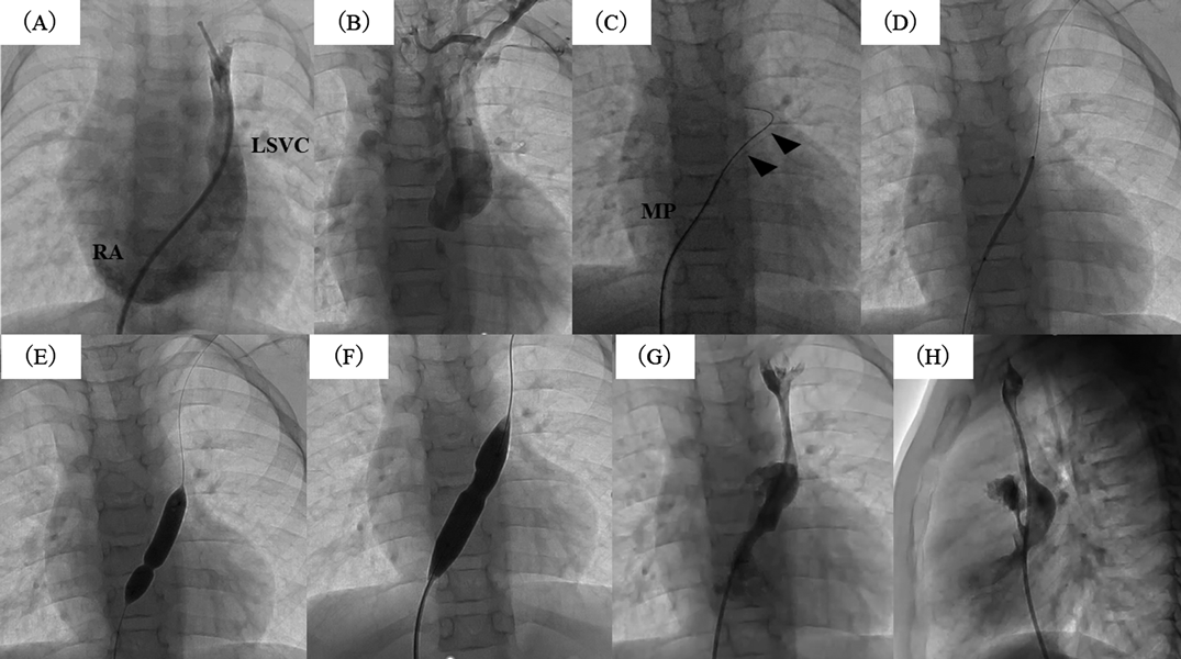
術後冠静脈洞閉鎖に対する経皮的バルーン血管形成術の一例Successful percutaneous balloon angioplasty of an acquired coronary sinus orifice atresia

1独立行政法人地域医療推進機構 九州病院小児科Department of Pediatrics, Kyushu Hospital, Japan Community Healthcare Organization ◇ Japan

2独立行政法人地域医療推進機構 九州病院心臓血管外科Department of Cardiovascular surgery, Kyushu Hospital, Japan Community Healthcare Organization ◇ Japan

受付日:2022年3月2日Received: March 2, 2022
受理日:2022年3月4日Accepted: March 4, 2022
発行日:2022年4月30日Published: April 30, 2022
HTMLPDFEPUB3

We report successful percutaneous balloon angioplasty of an acquired coronary sinus orifice atresia (CSOA) using a chronic total occlusion (CTO) guidewire via retrograde approach. A 5-year-old girl presented with facial edema. Her past history includes partial atrioventricular septal defect and left superior vena cava (LSVC) with unroofed coronary sinus that were successfully repaired by surgery at 7 months of age. Left-hand peripheral venography showed CSOA and congestion in the LSVC. Though we tried to insert multipurpose catheter into the coronary sinus with a 0.035″ guidewire via antegrade approach, this was not success. Therefore, we choose a 0.012″ tip CTO guidewire to penetrate acquired CSOA via retrograde approach and could successfully recanalize the pathway to the LSVC by stepwise dilation with different sized balloons. Contrast echocardiography after treatment confirmed that good patency from LSVC to RA.

Key words: acquired coronary sinus orifice atresia; CTO guide wire

症例

5歳女児.出生後に部分型房室中隔欠損症,冠静脈洞欠損を伴う左上大静脈(LSVC)と診断した.肺体血流比3.5,肺動脈圧31/29(19)mmHgであり,生後7か月時に一期的心内修復術を行った.LSVCは開口部をカットバックし下縁までV字型に降りるように縫合ラインを取り,右房(RA)へ流入するようにした.術後1か月後のLSVC造影でRAまでの流出を確認した(Fig. 1A).術後5年で顔面浮腫が出現し,造影CTでLSVC開口部狭窄を認めた.左上肢からの静脈造影で,LSVC内造影剤鬱滞を認め,RAは描出しなかった(Fig. 1B).後天的冠静脈洞閉鎖に経皮的バルーン血管形成術の方針とした.大腿静脈から逆行性に椎体をメルクマールとし,冠静脈洞口にマルチパーパスカテーテルを留置して0.035インチラジフォーカスガイドワイヤー(テルモ,東京)で穿通を試みたが困難だった.このため0.012インチ先端高加重ガイドワイヤー(ASAHI Conquest Pro,朝日インテック,愛知)を使用し穿通可能であった.SHIDEN® 2 mm(カネカメディックス,大阪),次にSterling® 8 mm(Boston Scientific; Marlborough, MA, USA),最後にMustang® 12 mm(Boston Scientific; Marlborough, MA, USA)で拡張し,LSVCの再疎通が可能だった(Fig. 1C–H).治療半年後のコントラストエコーで良好に開存を維持していた.

Journal of JCIC 6(2): 29 (2022)

Fig. 1 The patency from LSVC to RA was confirmed by postoperative angiography (A). The left-hand peripheral venography showed congested LSVC (B) and acquired coronary sinus orifice atresia. The 6F multipurpose using 0.014″ chronic total occlusion guidewire (arrowheads) successfully passed through into LSVC (C). Sequential balloon dilation using SHIDEN® (2×40 mm) (D), Sterling® (8×40 mm) (E), and Mustang® (12×40 mm) (F) successfully enlarged atretic coronary sinus orifice. Final angiography confirmed the patency from LSVC to RA in both anterior (G) and lateral view (H). Abbreviations LSVC, left superior vena cava; RA, right atrium

考察

既存の報告ではLSVCから冠状静脈洞まで順行性に冠静脈洞閉鎖部分をRFワイヤーで穿通している1–3)が,本症例では大腿静脈からの逆行性アプローチで先端高加重ガイドワイヤーを使用し,安全に術後性冠静脈洞閉鎖部穿通が可能であった.

この論文の要旨の一部は,第32回日本先天性心疾患インターベンション学会学術集会(2022年1月,岡山)において発表した.

This page was created on 2022-03-14T13:05:45.047+09:00
This page was last modified on 2022-04-25T14:00:31.000+09:00


このサイトは(株)国際文献社によって運用されています。