Journal of JCIC

Online edition: ISSN 2432–2342
JCIC学会事務局 JCIC学会事務局
〒162-0801東京都新宿区山吹町358-5アカデミーセンター Academy Center, 358-5 Yamabuki-cho, Shinju-ku, Tokyo 162-0801, Japan
Journal of JCIC 4(2): 18-23 (2019)
doi:10.20599/jjcic.4.18

原著原著

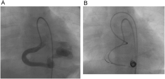
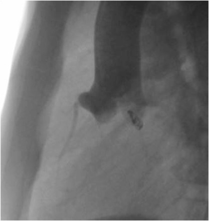
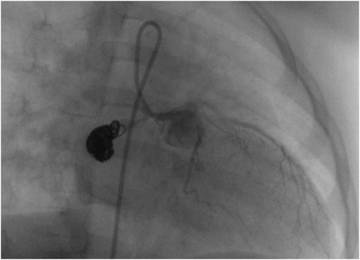
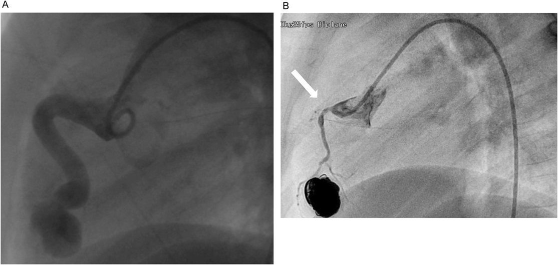
冠動脈瘻に対しカテーテル治療を行った症例の長期予後に対する検討Long-term Prognosis of the Coronary Arterial Fistulae treated by catheter intervention

神奈川県立こども医療センター循環器内科Department of Cardiology, Kanagawa Children’s Medical Center

受付日:2019年5月15日Received: May 15, 2019
受理日:2020年1月31日Accepted: January 31, 2020
発行日:2020年3月31日Published: March 31, 2020
HTMLPDFEPUB3

目的:冠動脈瘻(coronary arterial fistula: CAF)に対するカテーテル治療の長期的な予後を検討すること.

方法:対象は当院にてCAFに対してカテーテル治療を行った7例(年齢中央値1歳2か月)で,経過を後方視的に検討した.観察期間中央値は8年2か月(3か月~22年).治療法について検討するとともに,残存短絡の有無と冠動脈の形態について冠動脈造影により評価した.

結果:CAFは遠位型4例,近位型3例であった.全例,冠動脈の末梢側に各種デバイスを留置しCAFを閉塞した.中央値1年2ヶ月後と5年7ヶ月後に冠動脈造影を施行し,全例CAFは完全閉塞し,血栓形成や閉塞も認めなかった.形態については,正常化4例,caliber change 2例,盲端1例であり,4例では,抗血小板剤や抗凝固薬の内服を継続している.

結論:CAFに対するカテーテル治療の長期予後は良好である.冠動脈拡張を残す症例や狭窄病変を認める症例には,抗血小板剤や抗凝固薬の内服の継続を行う必要がある.

Objectives: The purpose of this study was to examine the long-term prognosis of coronary arterial fistula (CAF) treated by catheter intervention.

Method: Seven patients (median age 1.1 year) were enrolled who underwent catheter treatment for CAF in our institution. All cases were examined retrospectively. The median observational period was 8.1 years (minimum 3 months, maximum 22 years). Residual shunt and coronary morphology were examined by the selective coronary angiography.

Results: All patients achieved complete occlusion of CAFs. Follow-up coronary angiography showed no thrombus, nor obstruction. The morphology of coronary artery was normalized in 4 cases, revealed caliber change in 2 and blind pouch in one. Antiplatelet or anticoagulant were continued in 4 patients. The CAF was classified as distal types in 4 and proximal types in 3, and all cases were closed with various devices in the distal portion. Follow-up coronary angiography was performed in each after median 1.1 year and 5.5 years, and neither residual shunt, nor thrombus nor obstruction was recognized.

Conclusions: The long-term prognosis of catheter treatment for CAF is good. Follow-up catheterization is mandatory, and in cases where coronary artery dilatation remains or where stenosis is confirmed, the oral administration of antiplatelet agents and anticoagulants are required

Key words: Coronary artery fistula; transcatheter closure; Amplatzer Vascular Plug; antiplatelet agent; anticoagulant

はじめに

先天性冠動脈瘻(coronary arterial fistula;以下CAF)は稀な疾患で,有病率は0.2–0.6%と言われている1).短絡量が少量のものは自然閉鎖が期待出来るが2),中等度から大きな短絡量のあるものは心不全や心筋虚血,感染性心内膜炎,瘤の破裂のリスクを有する3).短絡量が少量のCAFに関する管理はまだ確立されていないが,中等度以上の短絡があるものは,一般的には治療の対象とされている.アメリカ心臓協会(American Heart Association;以下AHA)のガイドラインでは,有症状のCAFと,無症状でも大きな短絡のあるCAFに対しての閉鎖治療を推奨している4)

治療の方法としては,外科治療とカテーテル治療の2つがあるが,当院では解剖学的にデバイスでの閉鎖の方法論が成立すれば手術よりカテーテル治療を優先している.

対象と方法

対象は1996年~2018年に神奈川県立こども医療センターでCAFに対して,カテーテル治療を行った患者7人.CAFの大きさ,カテーテル治療の方法,塞栓後の経過を,診療録を用いて後方視的に検討した.患者の特徴をTable 1に示す.CAF発見の契機は,1例が胎児診断で,残り6例は心雑音であった.出生後の診断は中央値1か月(3日~4か月)と比較的早い段階でなされており,カテーテル治療施行時の年齢は中央値1歳2か月(4か月~3歳2か月)であった.全ての症例で心不全症状を認めなかった.心電図所見では,全例ST変化やT波異常を認めなかった.胸部レントゲン像では心胸郭比の平均は52%(47–59%)であった.経胸壁心臓超音波検査では全例著明に拡張した冠動脈を認め,左室拡張末期径の正常予測値に対する平均の割合は110%(83–130%)と容量負荷を認めた.CAFの拡張部の最大径は7–11 mm.起始部は右冠動脈が5例,左回旋枝が1例,左主幹部が1例で,開口部は,右房が3例,右室が2例,左室が2例であった.CAFと正常冠動脈の関係は近位型が3例,遠位型が4例であった.

Table 1 
PatientYear of procedureQp/QsAge of procedureFeeding coronary arterySite of drainageLargest diameter (mm)Type of CAF
119961.63y2mRCARA7.4proximal
220041.42y8mLCXRA(2 site)7proximal
320064mRCALV11.6distal
420108mLMTRA7.2proximal
520151.21y2mRCARV10.5distal
620161.01y9mRCALV10.5distal
720181.39mRCARV7.5distal
Qp/Qs, pulmonary to systemic flow ratio; CAF, coronary artery fistulae; RCA, right coronary artery; LMT, left main trunk; LCX, left 7circumflex coronary artery; RA, right atrium; LV, left ventricle; RV, right ventricle

2006年以降はバルーン付のカテーテルを併用しCAF内で,閉塞試験を施行した.さらに,バルーンを膨らませることにより,CAF内の血流を制限しコイル塞栓を行うflow control method (Fig. 1)を用いている.

Journal of JCIC 4(2): 18-23 (2019)

Fig. 1 The angiography of patient 3. The coronary artery fistula drained into the left ventricle (A). Flow-control method is used for the right coronary artery (RCA). One catheter is for coil implantation, and the other is for balloon inflation (B)

結果

1例は短絡部が2か所あり,1か所は閉塞し得たが,残りの1か所はコイル塞栓が困難で断念した.その他6例は目的とする短絡部にコイルまたは閉鎖栓を留置することが出来た.全例,治療後に冠動脈造影を行い,コイルや閉鎖栓の位置や他の血管からCAFへの血流供給がないかを確認した.2例はごく少量のシャントの残存を認めたが,5例は完全閉塞を確認した.コイルもしくはデバイスは全例,正常冠動脈から遠位で開口部近傍の可能な限りの遠位部に留置した.塞栓用デバイスとして1例にAmplatzer Vascular Plug II(AVP; Abbott Vascular, Santa Clara, California)を使用し,6例は着脱式のコイルを使用した.初期は,動脈管開存塞栓用着脱式コイルであるJackson detachableコイルや動脈管開存閉鎖用着脱式コイルであるFlipperコイルを使用していたが,近年では比較的柔らかいORBIT GALAXY(Depuy Synthes, USA)やTornado(Cook, Europe)を使用していた.重篤な合併症を認めなかったが,コイル塞栓中に心電図上,一過性にST上昇を来した症例を1例認めた.

フォローアップの血管造影検査は全例で行っており,2回行ったのは3例,1回行ったのが4例であった.フォローアップの血管造影検査の術後からの期間の中央値は4年11カ月(1年~9年10か月)で,全例短絡の残存を認めず,いずれの例も血栓形成や明確な虚血を示唆する狭窄や血流遅延といった所見は認めていない.

初期の2例のうち1例(症例1)はコイル留置後にパナルジンの内服を開始したが,フォローアップの血管造影検査(Fig. 2)を行い内服終了している.もう1例(症例2)は治療後,抗血小板剤も抗凝固剤も内服していなかった.中期2例では,コイル留置後アスピリンの内服を開始し,1例(症例3)はフォローアップの血管造影で冠動脈の正常化を確認しアスピリンの内服を中止し,もう1例(症例4)はCAFが近位型でフォローアップの血管造影検査でblind pouch(Fig. 3)を認め,アスピリンを継続した.後期3例のうち2例(症例5;Fig. 4),(症例6;Fig. 5)は拡張部の退縮が進行せず造影剤のwash-outに停滞を認める部位が残存するcaliber changeを呈したためアスピリンとワルファリンの内服を継続し,冠動脈の正常化を確認した1例(症例7)はワルファリンの内服は中止し,アスピリンは継続している.

Journal of JCIC 4(2): 18-23 (2019)

Fig. 2 The angiography of patient 1, which is the right coronary artery (RCA), performed 10 year after coil implantation

Journal of JCIC 4(2): 18-23 (2019)

Fig. 3 The angiography of patient 4, which is the left main trunk (LMT), performed 1 year after coil implantation. A blind pouch was observed between the coil and the LMT

Journal of JCIC 4(2): 18-23 (2019)

Fig. 4 The angiography of patient 5. The coronary artery fistula consisted drained into the right ventricle (A). The angiography of the RCA was performed 1 year after coil implantation. Caliber change (white arrow) was observed near the orifice of the RCA (B). The diameter of the coronary artery inflow potion was narrowed from 6 mm to 1.3 mm

Journal of JCIC 4(2): 18-23 (2019)

Fig. 5 The angiography of patient 6. The coronary artery fistula consisted drained into the left ventricle (A). The angiography of the RCA was performed 1 year after AVPII implantation. Caliber change (white arrow) was observed near the orifice of the RCA (B). The diameter of the coronary artery inflow potion was narrowed from 6 mm to 2.5 mm

Table 2 
PatientDevicesFlow stop methodResidual shuntComplicationDevice positions
1Flipper coil®5 mm×2+distal
2Flipper coil®5 mm×5+distal
3soft standard5 mm×12 mm×1+distal
5 mm×7 mm×2
Tornado®2 mm×4 mm×1
4Tornado®8 mm×20 cm×1+distal
5 mm×12 cm×6
5PRESIDIO® 18 cerecyte9 mm×33 mm×2++ST change (transient)distal
8 mm×24 mm×1
ORBIT GALAXY®7 mm×21 mm×2
6 mm×15 mm×2
6Amplatzer Vascular Plug®+distal
7ORBIT GALAXY®9 mm×25 mm×3+distal
8 mm×24 mm×1
7 mm×15 mm×1
Table 3 
PatientAge of follow-up cath①State of coronary arteryAge of follow-up cath②State of coronary arteryMedications
16y7mnormal13y1mnormalTiclopidine→stopped
27y7mnormal
31y1mnormal5y8mnormalAspirin→stopped after follow-up cath①
41y10mBlind pouch +6y4mBlind pouch+Aspirin continued
52y3mdilatationAspirin and Warfarin continued
Caliber change
63y2mdilatationAspirin and Warfarin continued
Caliber change
72ynormalWarfarin stopped Aspirin continued after follow-up cath①

考察

適応 AHAのガイドラインでは,有症状のCAFに対して閉鎖治療はClass I,症状はなくても大きな短絡のあるCAFに対しての閉鎖治療はClass IIaとしている4)

また,年齢が経ってからカテーテル治療の高い死亡率等を考慮すると,小児期に閉鎖しておく事が推奨されている5, 6).当院でも,心不全症状がなくても冠動脈の有意な拡張を来している大きな短絡の場合に治療対象としている.

治療方法の選択 治療方法としては,外科的にCAFを閉鎖する方法とカテーテルで塞栓する方法が挙げられるが,当院ではカテーテル治療を優先して行っている.外科手術よりもカテーテル治療を選択する理由に,外科手術後のほうが,合併症が多いとの報告がある1, 7).CAF閉鎖後の血栓形成の機序は不明ではあるが,CAFの血流減少が一因と考えられる.カテーテル治療でもカバードステントによる閉鎖例で治療翌日に血栓形成を認めた報告や6),外科的に閉鎖した例よりも,側副血行路の発達が少なかったとの報告もある5)ことより,急激な血流の変化が血栓の形成に寄与している可能性がある.一方で経皮的な治療による閉鎖では,徐々に冠動脈の縮小が得られ,冠動脈の血流の連続的な流れが持続し,いわゆる川崎病の冠動脈瘤が徐々に退縮する病態に近いのではないかと推測される.当院におけるカテーテル治療例での合併症は認めていない.

近位型では現在は,proximalとdistal両方に閉鎖栓をおくことが提唱されている5).Blind pouch盲端を避けるためにCAFの可能な限り正常冠動脈の近傍でコイルを塞栓するべきとする報告もあるが8),一方で近位部に留置する場合,コイルの突出による冠動脈狭窄から虚血を惹起するリスクによる留置困難例も見られる9).至適部位に限局的な狭窄部があれば,コイル留置は容易となる10).一方,遠位型のCAFに関しては,CAFの近位の正常冠灌流を阻害させないために可能な範囲でCAFに近接させて,デバイスないしコイルを留置する必要がある1)

血栓予防 CAFの治療後の血栓閉塞のリスクとしては冠静脈洞に還流している例や遠位型で冠動脈拡張が著しい例,近位型で異常血管遠位部を塞栓し盲端形成を生じた場合等に報告されている1, 8).症例4では,盲端となっているため注意深い観察が必要である.症例1は1度目のフォローアップの血管造影で,冠動脈の形態は概ね正常の形態をしており,狭窄はないためパナルジンを中止した.2回目のフォローアップの血管造影でも同様の所見であった.最近の当院の方針としては,カテーテル治療後には全例,アスピリンとワーファリンを投与し,治療して約1年後にフォローアップの血管造影を行い,拡張が退縮して(完全に正常レベルではなくても),造影剤の流れが良くなっていればワルファリンは中止しアスピリンは継続し,さらに5~6年後にフォローアップの血管造影を行い,内服の中止を検討するのがいいのではないかと考えている.そのため,症例7では1回目のカテーテルで冠動脈は正常化していたが,アスピリンは継続している.

また,早い時期,特に小児期での治療は更なるCAFによる異常血管の拡張を回避し,結果的に治療後の血栓のリスクを減らすことが出来る1)とする報告もあり,低年齢で治療することは,将来的な血栓形成のリスク軽減に寄与すると考える.

結語

7例のCAFに対して,コイル塞栓もしくはデバイス閉鎖を施行した.全例,重篤な合併症なく経過しており,CAFに対するカテーテル治療は安全で低侵襲な治療といえる.しかし,遠隔期で冠動脈拡張の残存や狭窄を認める症例が存在し,定期的なフォローアップの血管造影検査が必須であり,それに基づく適切な薬物療法が良好な転帰を獲得するための重要な要因と考えられた.

引用文献References

1) Gowda ST, Latson LA, Kutty S, et al: Intermediate to long-term outcome following congenital coronary artery fistulae closure with focus on thrombus formation. Am J Cardiol 2011; 107: 302–308

2) Schleich JM, Rey C, Gewillig M, et al: Spontaneous closure of congenital coronary artery fistulas. Heart 2001; 85: E6

3) Sunder KR, Balakrishnan KG, Tharakan JA, et al: Coronary artery fistula in children and adults: a review of 25 cases with long-term observations. Int J Cardiol 1997; 58: 47–53

4) Feltes TF, Becha E, Beekman RH 3rd, et al: Indication for cardiac catheterization and intervention in pediatric cardiac disease: a scientific statement from the American Heart Association. Circulation 2011; 123: 2607–2652

5) Thakkar B, Patel N, Poptani V, et al: Clinical and angiographic follow-up of coronary artery fistula interventions in children: techniques and classification revisited. Cardiol Young 2015; 25: 670–680

6) Jama A, Barsoum M, Bjarnason H, et al: Percutaneous closure of congenital coronary artery fistulae: results and angiographic follow-up. JACC Cardiovasc Interv 2011; 4: 814–821

7) Abdelmoneim SS, Mookadam F, Moustafa S, et al: Coronary Artery Fistula: Single-Center Experience Spanning 17 Years. J Interv Cardiol 2007; 20: 265–274

8) Valente AM, Lock JE, Gauvreau K, et al: Predictors of long-term adverse outcomes in patients with congenital coronary artery fistulae. Circ Cardiovasc Interv 2010; 3: 134–139

9) Kung GC, Moore P, McElhinney DB, et al: Retrograde transcatheter coil embolization of congenital coronary artery fistulas in infants and young children. Pediatr Cardiol 2003; 24: 448–453

10) 伊藤晋一,須田憲治,籠手田雄介,他冠動脈瘻に対するコイル塞栓術の検討.日本小児循環器学会雑誌2006; 22(5): 531–38

This page was created on 2020-02-18T09:47:38.761+09:00
This page was last modified on 2020-03-31T10:46:48.000+09:00


このサイトは(株)国際文献社によって運用されています。